Cytokeratin 5 Mouse Monoclonal Antibody
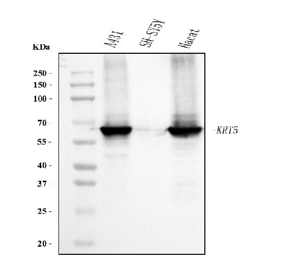
Western blot analysis of Cytokeratin 5 using anti- Cytokeratin 5 antibody. Electrophoresis was performed on a 5-20% SDS-PAGE gel at70V (Stacking gel) / 90V (Resolving gel) for 2-3 hours. The sample well of each lane was loaded with 30 ug of sampleunder reducing conditions. Lane 1: human A431 whole cell lysates Lane 2: human SH-SY5Y whole cell lysates Lane 3: human Hacat whole cell lysates. After electrophoresis, proteins were transferred to a nitrocellulose membrane at 150 mA for 50-90 minutes. Blocked the membrane with 5% non-fat milk/TBS for 1.5 hour at RT. The membrane was incubated with mouse anti- Cytokeratin 5 antigen affinity purified monoclonal antibody at 0.5 ug/mL overnight at 4°C, then washed with TBS-0.1%Tween 3 times with 5 minutes each and probed with a goat anti-mouse IgG-HRP secondary antibody at a dilution of 1:10000 for 1.5 hour at RT. The signal is developed using an Enhanced Chemiluminescent detection (ECL) kit with Tanon 5200 system. A specific band was detected for Cytokeratin 5 at approximately 62 kDa. The expected band size for Cytokeratin 5 is at 62 kDa.
| Catalog Number: | MAB-94762 |
| Conjugate: | Unconjugated |
| Size: | 200 ug |
| Concentration: | 1mg/ml |
| Host: | Mouse |
| Isotype: | IgG2b |
| Clone: | 5D3FT |
| Immunogen: | A synthetic peptide corresponding to a sequence in the middle region of human Cytokeratin 5 (286-317aa KVELEAKVDALMDEINFMKMFFDAELSQMQTH), different from the related mouse sequence by one amino acid, and identical to the related rat sequence. |
| Reactivity: | Human |
| Applications: | Western blot, 0.5-1 ug/ml Immunohistochemistry (Paraffin-embedded Section): 4-10 ug/ml Immunocytochemistry: 10 ug/ml Immunofluorescence,: 10 ug/ml Immunofluorescence, 10 ug/ml Flow Cytometry: 1-3 ug/1x106 cells |
| Molecular Weight: | 62 kDa |
| Purification: | Immunogen affinity purified. |
| Form: | Lyophilized: Add 0.2ml distilled water to obtain a concentration of 1mg/ml |
Applications
Western blot, 0.5-1 ug/ml Immunohistochemistry (Paraffin-embedded Section): 4-10 ug/ml Immunocytochemistry: 10 ug/ml Immunofluorescence,: 10 ug/ml Immunofluorescence, 10 ug/ml Flow Cytometry: 1-3 ug/1x106 cells
Immunogen
A synthetic peptide corresponding to a sequence in the middle region of human Cytokeratin 5 (286-317aa KVELEAKVDALMDEINFMKMFFDAELSQMQTH), different from the related mouse sequence by one amino acid, and identical to the related rat sequence.