GPX1
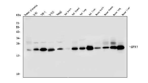
Western blot analysis of GPX1 using anti-GPX1 Antibody. Electrophoresis was performed on a 5-20% SDS-PAGE gel at 70V (Stacking gel) / 90V (Resolving gel) for 2-3 hours. The sample well of each lane was loaded with 50ug of sample under reducing conditions. Lane 1: human placenta tissue lysates, Lane 2: human U-87 whole cell lysates, Lane 3: human THP-1 whole cell lysates, Lane 4: human U-937 whole cell lysates, Lane 5: human HepG2 whole cell lysates, Lane 6: rat brain tissue lysates, Lane 7: rat thymus tissue lysates, Lane 8: rat lung tissue lysates, Lane 9: rat liver tissue lysates, Lane 10: mouse brain tissue lysates, Lane 11: mouse thymus tissue lysates, Lane 12: mouse lung tissue lysates, Lane 13: mouse liver tissue lysates. After Electrophoresis, proteins were transferred to a Nitrocellulose membrane at 150mA for 50-90 minutes. Blocked the membrane with 5% Non-fat Milk/ TBS for 1.5 hour at RT. The membrane was incubated with mouse anti-GPX1 antigen affinity purified monoclonal antibody at 0.5 ug/mL overnight at 4°C, then washed with TBS-0.1%Tween 3 times with 5 minutes each and probed with a goat anti-mouse IgG-HRP secondary antibody at a dilution of 1:10000 for 1.5 hour at RT. The signal is developed using an Enhanced Chemiluminescent detection (ECL) kit with Tanon 5200 system. A specific band was detected for GPX1 at approximately 22KD. The expected band size for GPX1 is at 22KD.
| Catalog Number: | MAB-94602 |
| Size: | 100μg |
| Concentration: | 1mg/ml |
| Host: | Ms |
| Isotype: | IgG |
| Clone: | 8B10 |
| Immunogen: | A synthetic peptide corresponding to a sequence in the middle region of human GPX1 (116-146aa EVNGAGAHPLFAFLREALPAPSDDATALMTD), different from the related mouse sequence by six amino acids and from the related rat sequence by five amino acids. |
| Reactivity: | Hu, Ms, Rt |
| Applications: | Western blot: 0.2-1ug/ml Immunohistochemistry(Paraffin-embedded Section): 1-2ug/ml Flow Cytometry: 1-3ug/1x106 cells |
| Purification: | Aff. Pur. |
| Form: | Liquid |
Applications
Western blot: 0.2-1ug/ml Immunohistochemistry(Paraffin-embedded Section): 1-2ug/ml Flow Cytometry: 1-3ug/1x106 cells
Immunogen
A synthetic peptide corresponding to a sequence in the middle region of human GPX1 (116-146aa EVNGAGAHPLFAFLREALPAPSDDATALMTD), different from the related mouse sequence by six amino acids and from the related rat sequence by five amino acids.