Paxillin Mouse Monoclonal Antibody
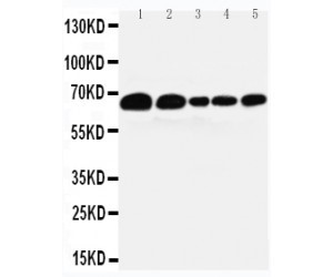
Western blot analysis of Paxillin using anti-Paxillin antibody. Electrophoresis was performed on a 5-20% SDS-PAGE gel at 70V (Stacking gel) / 90V (Resolving gel) for 2-3 hours. The sample well of each lane was loaded with 30 ug of sample under reducing conditions. Lane 1: human A431 whole cell lysates Lane 2: human Hela whole cell lysates Lane 3: human MCF-7 whole cell lysates Lane 4: human CACO-2 whole cell lysates Lane 5: rat liver tissue lysates Lane 6: rat RH35 whole cell lysates Lane 7: mouse liver tissue lysates. After electrophoresis, proteins were transferred to a nitrocellulose membrane at 150 mA for 50-90 minutes.Blocked the membrane with 5% non-fat milk/TBS for 1.5 hour at RT. The membrane was incubated with mouse anti-Paxillin antigen affinity purified monoclonal antibody at 1 ug/mL overnight at 4°C, then washed with TBS-0.1%Tween 3 times with 5 minutes each and probed with a goat anti-mouse IgG-HRP secondary antibody at a dilution of 1:10000 for 1.5 hour at RT. The signal is developed using an Enhanced Chemiluminescent detection (ECL) kit with Tanon 5200 system. A specific band was detected for Paxillin at approximately 65 kDa. The expected band size for Paxillin is at 65 kDa.
| Catalog Number: | MAB-80123 |
| Conjugate: | Unconjugated |
| Size: | 100 ug |
| Concentration: | Adding 500ul of PBS buffer will yield a concentration of 100 ug/500ul. |
| Host: | Mouse |
| Isotype: | IgG1 |
| Clone: | PXC-10 |
| Immunogen: | C-terminal part of recombinant chicken paxillin (amino acids 305-559). |
| Reactivity: | Bovine, Chicken, Hamster, Human, Mouse, Rat |
| Applications: | Immunocytochemistry: 1ug/ml Western blot: 1-2ug/ml |
| Molecular Weight: | 65kDa |
| Purification: | Ascites |
| Form: | Liquid |
Applications
Immunocytochemistry: 1ug/ml Western blot: 1-2ug/ml
Immunogen
C-terminal part of recombinant chicken paxillin (amino acids 305-559).