SETD1A
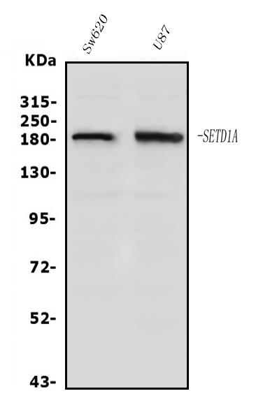
Western blot analysis of hSET1/SET1/SETD1A using anti-hSET1/SET1/SETD1A antibody. Electrophoresis was performed on a 5-20% SDS-PAGE gel at70V (Stacking gel) / 90V (Resolving gel) for 2-3 hours. The sample well of each lane was loaded with 50ug of sample under reducing conditions. Lane 1: human SW620 whole cell lysates, Lane 2: human U87 whole cell lysates. After Electrophoresis, proteins were transferred to a Nitrocellulose membrane at 150mA for 50-90 minutes. Blocked the membrane with 5% Non-fat Milk/ TBS for 1.5hour at RT. The membrane was incubated with rabbit tihSET1/SET1/SETD1A antigen affinity purified polyclonal antibody at 0.5 ug/mL overnight at 4°C, then washed with TBS-0.1%Tween 3 times with 5minutes each and probed with a goat anti-rabbit IgG-HRPsecondary antibody at a dilution of 1:5000 for 1.5 hour atRT. The signal is developed using an ECL West Pico Plus kit with Tanon 5200 system. A specific band was detected forhSET1/SET1/SETD1A at approximately 186KD. The expectedband size for hSET1/SET1/SETD1A is at 186KD.
| Catalog Number: | AB-84349 |
| Size: | 100ug |
| Concentration: | 1mg/ml |
| Host: | Rb |
| Isotype: | IgG |
| Clone: | POLY |
| Reactivity: | Hu, Ms, Rt |
| Applications: | Western Blot: 0.25-0.5ug/ml Immunohistochemistry (Paraffin-embedded Section):0.5-1ug/ml Immunofluorescence: 2ug/ml, Human ELISA: 0.1-0.5mug/ml |
| Molecular Weight: | 186kDa |
| Purification: | Aff. Pur. |
| Form: | Liquid |
Applications
Western Blot: 0.25-0.5ug/ml Immunohistochemistry (Paraffin-embedded Section):0.5-1ug/ml Immunofluorescence: 2ug/ml, Human ELISA: 0.1-0.5mug/ml